Palisade mesophyll cell
In brief
Palisade mesophyll cells belong to a leaf’s internal photosynthetic tissue. In a typical eudicot leaf they are relatively closely packed beneath the upper epidermis and contain chloroplasts. Nearby spongy mesophyll is more loosely arranged, with air spaces supporting gas exchange. Cell, tissue and organ remain distinct structural levels. [7]
Evidence for this statement
Source-based explanation
Where to check this statement
- biology59-leaves
- Structure of a Typical Leaf; Leaf Structure and Function; Leaf Adaptations
Save, investigate and check this version
Editorial draft · human review pending
Ask about this materialOn this page
A photosynthetic cell inside a leaf
What three-dimensional imaging adds
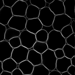
Wuyts and colleagues reconstructed Arabidopsis leaf tissues from stacks of optical sections. Their Results distinguish palisade cells from lobed spongy cells and show why one transverse view can confuse them. Several views preserve cellular context. This helps ask whether growth involves changes in cell number, dimensions or tissue organisation.
[2]Evidence for this statement
EcoGenesis
Where to check this statement
- photo-cell
- Results: Tissue level growth variables were reliably measured for all layers of mesophyll tissue; Figures 2 and 6
One cell type
This page describes a palisade mesophyll cell, not the whole mesophyll tissue. The identifier PO:0006206 preserves that distinction. Links to palisade tissue and leaf lead to larger structures while keeping each concept at its own level.
[1]What the optical section shows
The Wuyts and colleagues image shows stained cell walls. Its caption identifies Arabidopsis Col-4, leaf six and 19 days after initiation. These details belong to that material; they do not determine the age of every leaf shown elsewhere in the encyclopedia.
[2]Evidence for this statement
Explanation of retained sources
From cell to organelle
Chloroplast structure is illustrated by a separate electron micrograph with its own caption and scale. This changes source material rather than magnifying the same optical section. The PsbA record adds another level: annotation of a particular protein, not a measurement of its presence in the selected cell.
[2] [4] [5]Evidence for this statement
Explanation of retained sources
- Arabidopsis thaliana Col-4, leaf 6, 19 days after initiation; Figure 2a, mature palisade mesophyll
- Arabidopsis thaliana, first leaf of control seedling grown without added NaCl; Figure 3A
- UniProt P83755 · entry 161 / sequence 2
Where to check this statement
- uniprot-P83755
- Captured provider JSON; named selected record and native source versions
Connected concepts
Chloroplast
A chloroplast is a chlorophyll-containing plastid. It contains thylakoids and stroma, with some thylakoids organised into stacks called grana.
[3] [4] [5]Evidence for this statement
Source-based explanation
- GO:0009507 · chloroplast
- Arabidopsis thaliana, first leaf of control seedling grown without added NaCl; Figure 3A
- UniProt P83755 · entry 161 / sequence 2
Where to check this statement
- go-chloroplast
- Captured GO ontology JSON: results[].id and definition.text
- uniprot-P83755
- Captured provider JSON; named selected record and native source versions

Continue with an organism
The photographs and molecular record come from separate source materials.
Your research question
Save the article to return to your question and this source version.
Plan a test
This is your research plan. Completing these fields does not confirm a hypothesis.
Sources and versions
Editorial draft · human review pending
- Plant Ontology · 2026-01-09
2026-09-04 · CC-BY-4.0
Provenance and original
- Source version
- 2026-01-09 / f5e2a95d88fe08af22f49e522cc78481d990a0b0
- Arabidopsis thaliana Col-4, leaf 6, 19 days after initiation; Figure 2a, mature palisade mesophyll
Retrieval date unknown · CC BY 2.0
Provenance and original
- Original SHA-256
- 5510c5c1dba4162b182eed9ae054d2e97c25cc529740a5125ffcb1fd1c001b3c
- Processing
- Mature palisade panel extracted from Figure 2a; encoded as WebP. The shared scale bar is outside this panel and remains available in the full source figure.
- GO:0009507 · chloroplast
2026-09-09T01:17:34.931415+00:00 · CC-BY-4.0
Provenance and original
- Original SHA-256
- 13082607c7dcc9e92cd281f1fc6a8fdf2878bceba74e9d7b6ad50d43b0ce33ba
- Processing
- Unmodified provider response; article text is an attributed educational summary.
- Version details
- QuickGO response did not supply a native ontology release; retrieval time and exact response hash retained.
- Arabidopsis thaliana, first leaf of control seedling grown without added NaCl; Figure 3A
Retrieval date unknown · CC BY 4.0
Provenance and original
- Original SHA-256
- fe305c7d3a3327a06dda04c186989b5b8b6dec101bfe13aa851a3bc47dc07b14
- Processing
- Panel A extracted; original g/t annotations and 1 µm scale retained; encoded as WebP.
- UniProt P83755 · entry 161 / sequence 2
2026-09-09T01:17:34.554931+00:00 · CC-BY-4.0
Provenance and original
- Source version
- entry 161; sequence 2
- Original SHA-256
- f16a5f33fbf85a12aa5c91ab31f6092f0bbe3be874040c84c4954e87b20ae3a9
- Processing
- Unmodified provider response; article text is an attributed educational summary.
- Version details
- Provider entry/sequence versions retained.
- Arabidopsis thaliana basal leaf; photographed 14 April 2021
Retrieval date unknown · CC BY-SA 4.0
Provenance and original
- Original SHA-256
- 4c0da1e60a566728712e6443d8dfa11d4f4cd0084a0ef454d1262a3cdf05fa39
- Processing
- Resized and encoded as WebP; original 5 mm scale retained.
- Clark, Douglas & Choi · Biology 2e · 30.4 Leaves
2026-10-01 · Terms of use on source site
Provenance and original
- Source version
- Selected web passages checked 2026-10-01; no immutable response edition asserted
- Source reading scope
- Named background passages only; not new experimental evidence.
- Location in the original source
- Structure of a Typical Leaf; Leaf Structure and Function; Leaf Adaptations
Inspect structured data
- Content version
- e3f5527c624509f9cb96e4a38ea8b68d50bed5431eca7b4091d21ff28ae70463
- Source version
- knowledge-20260909.1
- Updated
- 2026-10-01